


X-rays
X-rays provide your healthcare team with valuable information to diagnose conditions throughout the body while limiting radiation with our digital X-ray technology.
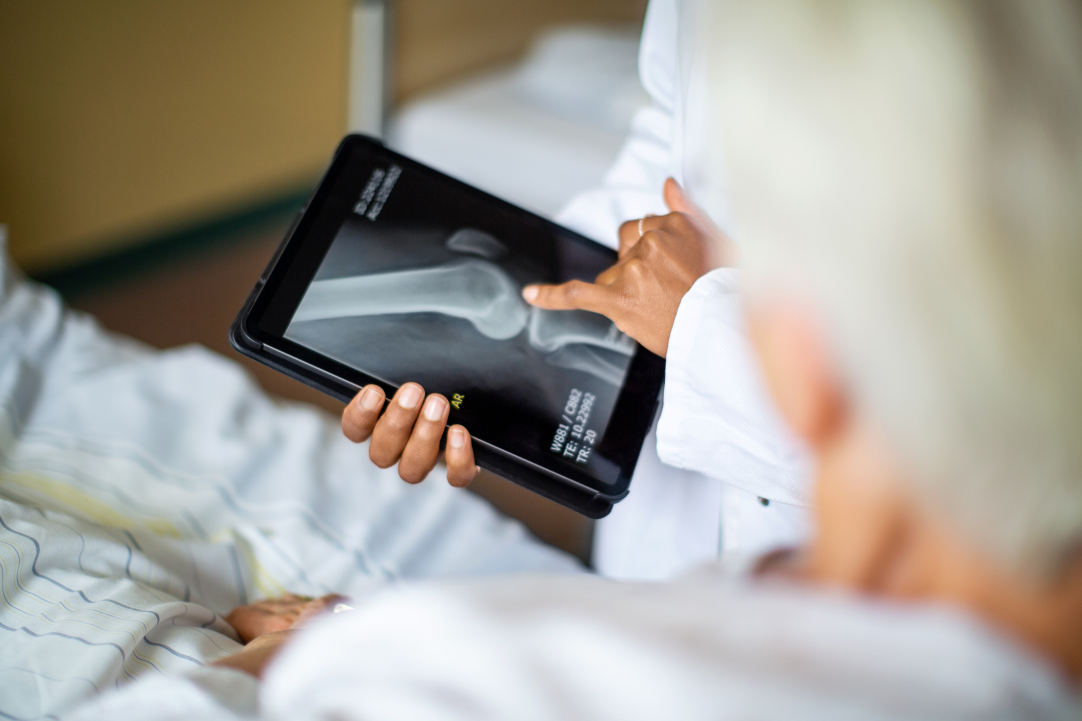
X-rays use a small dose of ionizing radiation to produce images. Calcium absorbs X-rays more than other parts of the body, so bones show up white. Fat and other soft tissues absorb less of the X-rays and look gray. Lungs look black because air absorbs the least amounts of X-rays.
X-rays evaluate:
- Abdominal and GI tract conditions
- Arthritis
- Bones and joints
- Blood vessels near your heart and the size of your heart
- Breast cancer (mammograms)
- Pneumonia (chest X-ray)
- Teeth
- Tumors
X-ray technologists will protect parts of your body from receiving unnecessary radiation by having you wear a lead apron.
Preparing for an X-ray exam
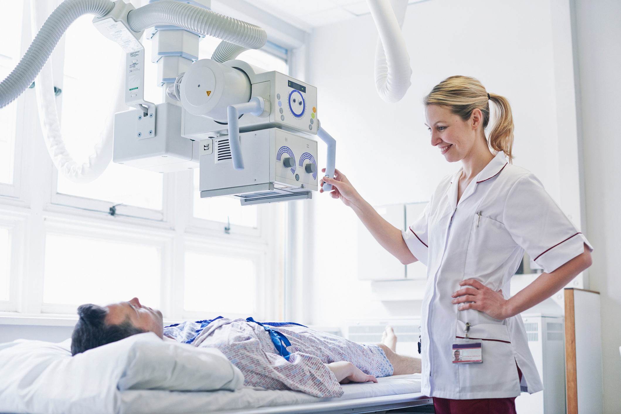
For most X-rays, you’ll be asked to put on a hospital gown. You may need to take off jewelry, glasses or anything else that could interfere with the image. You may be asked to lie on a table or stand up, depending on the part of the body being scanned.
At Spartanburg Regional Healthcare System, all our X-ray technologists are registered with the American Registry of Radiology Technologists. They are trained in administering the least amount of radiation possible to achieve the images your provider has requested.
X-rays are a noninvasive test that can be completed in 15 to 30 minutes, and most X-rays don’t require an appointment. For your convenience, we also offer Saturday hours at some locations.
Fluoroscopy at Spartanburg Regional
Fluoroscopy uses X-rays to produce a continuous image that’s like a video instead of a static picture. This technique allows your healthcare team to look at your body’s processes and organs in real time with the help of contrast dye.
The dye is administered orally, through an IV or with an enema. It helps radiologists see how an organ or system functions in real time.
Fluoroscopy helps doctors learn more about the skeletal, digestive, urinary, respiratory and reproductive systems by looking at:
- Bones and joints
- Heart function
- Kidneys
- Lungs
- Muscles
Fluoroscopy is also used during interventional radiology procedures to diagnose or treat conditions. Some uses include:
- Conducting biopsies
- Directing the movement of a catheter through blood vessels, bile ducts or the urinary system
- Performing angiograms to look at blood vessels and blood flow
- Placing devices within the body, such as stents that open blocked vessels
Your fluoroscopy exam
Because of the use of contrast dye, fluoroscopy takes longer than an X-ray. You will wear a hospital gown for the procedure and remove any jewelry or other items that would interfere.
After you receive the contrast agent, you’ll be positioned on the X-ray table to best view the area being studied. You may be asked to move a certain body part or hold your breath at intervals.
A fluoroscopy study can take up to an hour and an appointment is required.